print, photography, gelatin-silver-print
#
aged paper
#
homemade paper
#
paperlike
# print
#
sketch book
#
hardpaper
#
personal journal design
#
photography
#
personal sketchbook
#
journal
#
gelatin-silver-print
#
sketchbook drawing
#
sketchbook art
#
realism
Dimensions: height 127 mm, width 115 mm
Copyright: Rijks Museum: Open Domain
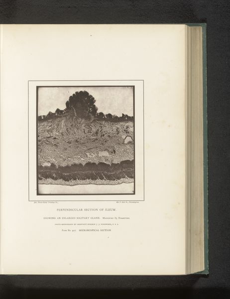
This is a microscopic photograph of a perpendicular section of a colon coated with pseudomembrane, by Joseph Janvier Woodward. The image offers a fascinating window into the intersection of art, science, and the institutional structures that supported their development in 19th century America. Woodward was a surgeon in the US Army Medical Corps during the Civil War. He pioneered the use of photomicrography - photography through a microscope - to study diseases. Published in medical journals and textbooks, Woodward’s images served a practical function for doctors and scientists. But they also reflect the broader cultural fascination with scientific progress and the power of the image. Institutions such as the Army Medical Museum played a vital role in this moment. Historians can use these images as primary sources in their study of the history of medicine, photography, and the development of scientific institutions in the 19th century. This allows us to interpret these images as culturally and historically contingent objects.
Comments
No comments
Be the first to comment and join the conversation on the ultimate creative platform.
More like this