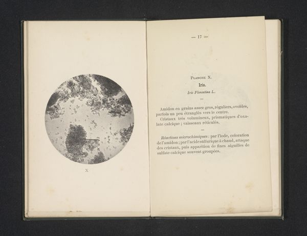
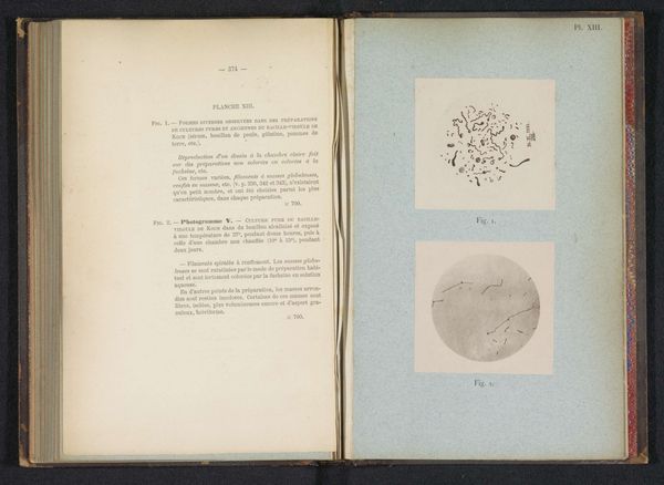
before 1885
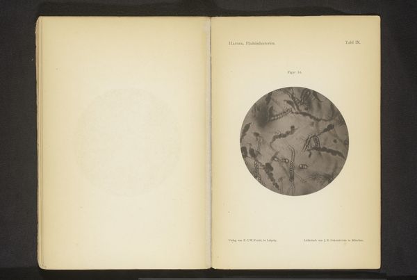
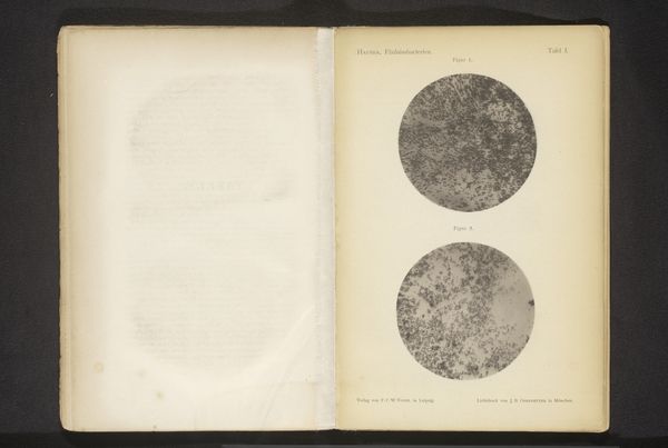
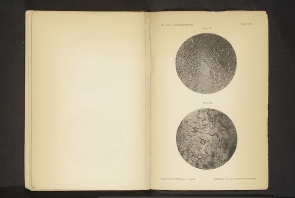
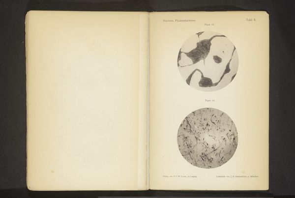
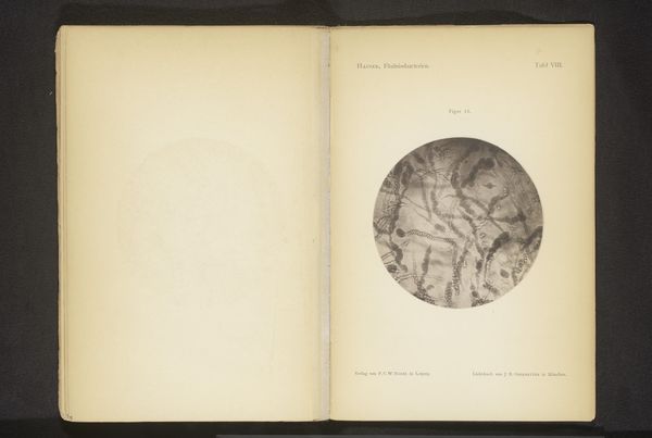
Twee microscoopopnamen van het darmslijmvlies van iemand die is gestorven aan cholera
Listen to curator's interpretation
Curatorial notes
Émile van Ermengem made these images using microscopy and photography, processes that blend scientific precision with visual representation. Van Ermengem captured these images of the intestinal lining of a cholera victim, revealing the disease's impact at a microscopic level. The photographs provide a stark depiction of cellular damage and bacterial presence, made visible through careful preparation and magnification techniques. The creation of such images required both technical skill in microscopy and photographic expertise to document the findings. These photographs also speak to the broader social context of the late 19th century, marked by significant advancements in bacteriology and public health. The study of infectious diseases like cholera prompted intensive scientific investigation and public health initiatives, reflecting the period's focus on understanding and combating widespread health crises. By examining the processes and context behind this image, we gain insight into the intersection of scientific inquiry, technological innovation, and social responsibility in addressing public health challenges.