#
pencil drawn
#
toned paper
#
light pencil work
#
pencil sketch
#
personal sketchbook
#
pencil drawing
#
portrait drawing
#
pencil work
#
watercolour illustration
#
pencil art
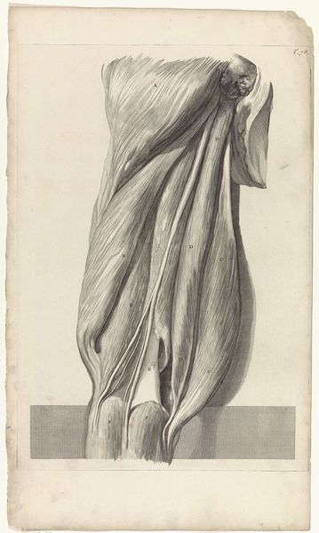
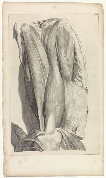
Dimensions: width 338 mm, height 490 mm
Copyright: Rijks Museum: Open Domain
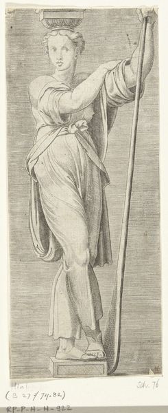
Pieter van Gunst created this anatomical study of the femur using etching and engraving techniques. Produced in the Netherlands, probably in the late 17th or early 18th century, the image reflects the growing scientific curiosity of the period. The Dutch Golden Age was a time of intense focus on observation, classification, and the pursuit of knowledge. Illustrated anatomical studies like this one served as important tools for medical education and research. The level of detail in the rendering is a testament to the importance placed on accurate representation in scientific inquiry. Of course, it’s worth remembering that the images depended on access to bodies, raising ethical questions about the sources of specimens. Understanding the history of science means considering the social and institutional conditions that make such images possible. Historians can use a range of resources such as medical texts, biographies of scientists, and institutional records to help us understand the role played by images such as this within the broader culture.
Comments
No comments
Be the first to comment and join the conversation on the ultimate creative platform.
More like this